
Uterus Definition, Shape, Size, Location and Function in Human Body
- What did the first Stone Age Man utter with his face to the sky?
- How did the ancient Greek scholars describe uterus?
- What is the uterus location and size?
- When isthe retroverted uterus said to be normal?
Did you know for many centuries both religion and law forbade the dissection of human bodies? Therefore, all the concepts about the anatomy of human reproductive tract were based on findings in animals.
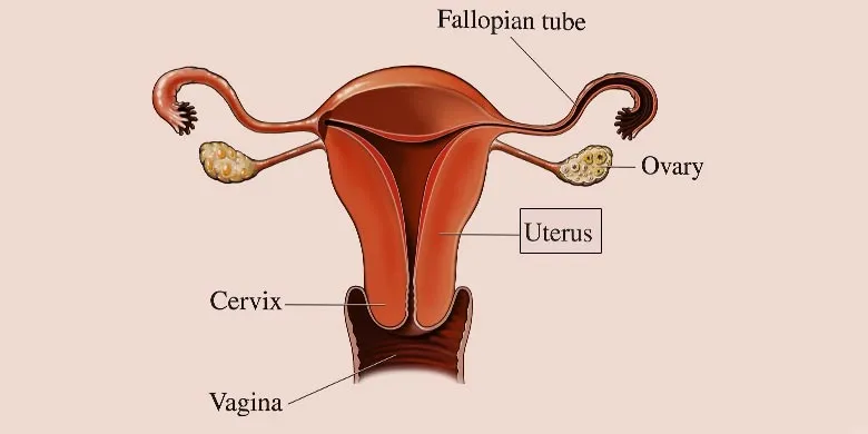
Fig. 1: Uterus
“The Uterus” edited be T. Chard and J. G. Grudzinskas (1994) begins with the question “Where did I come from?” purportedly uttered by the first Stone Age Man with his face raised to the sky. The very question has perplexed the intellectuals throughout the ages. Both theology and science have been investigating it in close collaboration with each other.
The Uterus in Historical Perspective:
The uterus in human body itself is a large and engrossing subject and our present understanding of it is the result of the philosophical and scientific endeavours undertaken during the past several centuries.
At the outset, in the 4th century BC, it was Hippocrates the great “Father of Medicine” who rejected the belief about the role of supernatural forces in causing or curing disease. His idea that diseases could be cured by correcting the imbalance of “humours” in the body paved the way for the study of patient’s body. In the nascent study of human anatomy and physiology, the uterus became an object of great attention.
Did you know? The uterus was initially described as a reproductive structure comprised of several cavities showing angulation and horns and their lining studded with suckers or tentacles.
Female Reproductive Organs:
The organs of the female reproductive system can be divided into the external genitalia and the internal genital organs. The external genitalia are in the diamond-shaped area below the pelvic diaphragm and between the legs, called perineum.
On the other hand, the internal genital organs are housed inside the pelvis except vagina, which lies partly in the pelvic region and partly in the perineum. The internal genital organs include the vagina, the uterus, the uterine or fallopian tubes, and the ovaries or female gonads.
Uterus Definition and Function:
A crucial organ in the female reproductive system, the uterus or womb can be defined as a hollow muscular organ placed in the female pelvis between the rectum and the urinary bladder. Made up of thick walls, it appears like an upside-down pear.
In the embryonic stage, the womb develops from the middle horizontal part and the proximal vertical part of the Mullerian duct (also called the paramesonephric duct) on each side.
The main job of the womb is to house and nourish the developing foetus until it is ready for birth. In addition to holding the baby during pregnancy, it also plays a key role in menstruation.
The Uterus vs the Uterine Tube:
Also called the fallopian tubes or oviducts, the uterine tubes are the female reproductive structures that serve as a passageway for the transport of ova from the ovary to the uterus. A uterine tube also acts as the site for the fertilization of the egg by a sperm while the uterus is the sight for the implantation of the fertilized egg.
Shape and Size:
In the book, “Clinical Anatomy: A Problem Solving Approach”, Neeta V. Kulkarni describes the uterus as a piriform or pear-shaped hollow, muscular organ measuring 7.5 x 5 x 2.5 cm in its dimensions of length, breadth and thickness, respectively. Here the fundus and the body run for the length of 5 cm while the remaining 2.5 cm is contributed by the cervix.
The Uterine Cavity:
The inside of the uterus is called the uterine cavity. This cavity is divisible into two major components, i.e. the cavity of the cervix or cervical canal and the cavity of the body. The cervical canal is spindle-shaped while the cavity of the body looks like the inverted isosceles triangle.
The internal orifice allows the cervical canal to communicate with the cavity of the body while the external orifice develops its link with the vagina. Therefore, it can be said that the cervical canal runs between the internal and external orifices.
When measured by an instrument called the uterine sound, the uterine cavity seems to be running for the length of 6 cm from the wall of the fundus to the external orifice. The ratio of the length of the cervix and of the uterus may vary with age. That is, from birth till the female reaches puberty, this ratio is 2:1 but after puberty it becomes 1:2.
Uterus Location and Position:
Although the uterus is situated between the rectum behind and urinary bladder in front in the pelvic cavity, its position and orientation may vary from individual to individual. For example, it may be anteverted or retroverted. In anteverted position, the womb tips slightly forward.
The position of the uterus is described as anteverted and anteflexed when the urinary bladder is empty. In this position, its body is tilted forward on urinary bladder’s superior surface which provides support.
Anteversion results from the angulation between the long axes of the vagina and that of the cervix. So, the uterus is bent forward at 90 degrees as the angle of version. However, a fully distended bladder pushes the uterus in line with the vagina, thus making its position retroverted. Such a retroverted position of the uterus is considered normal if it occurs due to the fully distended bladder.
Anteflexed uterus results from the angulation between the long axes of the vagina and the body of the uterus. This angle may measure between 120 and 170 degrees. This angulation bends the uterus downward.
Abnormal Uterine Positions:
Retroflexion and retroversion are said to be the abnormal positions of uterus. In case of retroflexion, there is no angulation between the cervix and uterus. Therefore, the uterus bends backwards on the cervix making the cervical canal narrower. Such a position is likely to minimize the possibility of conception.
Retroversion, on the other hand, causes the uterus to bend backwards in the rectouterine pouch. This position predisposes to the prolapse (slipping down or forward from its normal position) of the uterus.
Uterus Diagram and Parts:
In the uterus diagram, you will see that sitting in the pelvic cavity this hollow muscular structure communicates with the vagina inferiorly and with the uterine tube superiorly on each side.
A dissection of the uterine wall shows that it is made of three layers, namely, the inner endometrium, the middle myometrium, and the outer perimetrium. These constituent layers are also called mucosa, muscular coat, and serous coat, respectively.
Three major parts of the organ are clearly distinguishable in the uterus diagram. They are the fundus, the body and cervix.
The Fundus:
The rounded upper part just above the level of the entrance of uterine tubes is called the fundus.
The Body:
The body is that part of womb that lies between the fundus and the isthmus. Here the isthmus is the constricted part lying between the body and cervix of the uterus and measuring about half a centimetre.
The body has the anterior and posterior surfaces and the left and right margins. When the organ is in its normal anteverted or anteflexed position, the posterior surface faces upward and anterior downwards. On the other hand, the left and right lateral margins are related to the uterine artery and give attachment to the broad ligament. The superolateral angle or cornu of the uterus is the upper end of the lateral margin. Housing the tubo-uterine junction, this angle gives attachment to the ligament of uterus and ligament of ovary.
The Cervix:
It extends from the isthmus and ends at the external orifice, which opens into the vagina through a perforation in its anterior wall. So, the cervix is distinguishable into the vaginal and supravaginal parts.
Menstrual Cycle:
The ovarian hormones trigger cyclical changes in the endometrium or mucosa of the uterus, called menstrual cycle, which continue from menarche to menopause.
Uterus Diseases:
Several uterus diseases of mild and serious nature are known today. Most of them have effective cure if timely diagnosed and treated. Some pathological states of the womb include prolapse of the uterus, carcinoma of the cervix and of the uterus, fibroids, adenomyosis, endometritis, hematometra, and so on.
The prolapse of the uterus is a condition characterized by the descension of the uterus in the vagina. The prolapse may occur due to repeated childbirth, perineal tears, and the loss of tone of muscles of pelvic diaphragm. In extreme cases (in procidentia), the whole uterus may protrude outside the vaginal orifice.
About the Author