
Top 12 Heart Functions - Heart Location with Diagram
Pumping 2,000 gallons of blood with 100,000 beats daily, the heart tirelessly works till your last breath. 1 in 3 US women dies of heart disease! Larger hearts beat slower. How? Read on to discover fascinating facts about the function of the heart. If you know “What is the function of the heart?” it’ll help you ease its workload and prevent a disorder. Here are the primary and secondary heart functions in brief.
- Pulmonary Circulation :It carries the blood from the heart to the lungs for oxygenation.
- Systemic Circulation :Extending from the aorta, it pushes oxygen throughout the body.
- Coronary Circulatione :Your heart’s dedicated blood supply to take care of cardiac muscle tissue.
- Splanchnic Circulation :Your spleen, liver, and pancreas get nutrients through this.
- Cutaneous Circulation :It fulfills the skin’s relatively small requirements for oxygen and nutrients.
- Cerebral Circulation :Your brain gets 15-20% of cardiac output through this system.
- Pumping Interstitial Fluid The lymphatic system returns the excess interstitial fluid to the circulatory system.
- Generation of electrical impulse :The impulses initiate cardiac action to maintain life.
- Distribution of Nutrients :Digested food from the small intestine reaches every cell.
- Oxygen Supply :The most essential fuel, oxygen all the tissues ‘breathe’.
- Hormones Delivery :Hormones from extracellular fluid reach their target sites.
- Disposing of Wastes :Removes wastes, including CO2, the principal waste product.
In addition to elaborating on the heart function. The present article explains the human heart's structure, parts, diagram, location, and facts. Also, you’ll get answers to the following FAQs about the heart:
It is the main organ of your body's circulatory system, pumping blood throughout the body. Understand the heart's structure and function, it is a muscular pump that contracts at regular intervals to squeeze the blood into the blood vessels.
Top 12 Heart Functions in Detail:
1. Pulmonary Circulation:
The pulmonary component of the circulatory system begins at the junction between the right ventricle and the pulmonary artery. They purify the blood, i.e., restore its oxygen content and eliminate excess carbon dioxide. The blood that the superior and inferior vena cavae collect from the upper and lower halves of the body, respectively, is deficient in oxygen.
Numerous pulmonary capillaries diverged from the right and left pulmonary arteries that help the blood. They take up oxygen and unload carbon dioxide in the lungs. All this blood is fully saturated with oxygen and transported back to the heart via the pulmonary vein. The veins carrying oxygenated blood empties its contents into the left atrium.
The upper left chamber then pushes it into the left ventricle via the left atrioventricular (or mitral) valve. Oxygen-rich blood leaves the heart and enters the aorta through the aortic valve, which marks the starting point of the systemic circulation.
2. Systemic Circulation:
It starts at the aortic valve, where the aorta is the largest artery in the human body. It joins the left atrium and ends at the junction of the superior and inferior vena cavae with the right atrium. Its primary job is to supply the peripheral organs and tissue beds with oxygen-rich blood and return the venous or oxygen-depleted blood to the pumping organ.
Meanwhile, large and small arteries distribute the blood throughout the body, and a network of veins recollects and brings it back for oxygenation. The capillaries are the exchange vessels, serving as a connecting link between the arteries and veins. The main job of the capillaries is to facilitate the exchange of nutrients and fluid between the blood and the interstitial space.
Did you know?The systemic circulation contains nearly 80 percent of your body’s blood volume! Meanwhile, the pulmonary and coronary vascular circuits contain 9-12 and 8-11 percent of the total blood volume, respectively.
3. Coronary Circulation:
It begins at the coronary ostia, where the ascending aorta branches off, and ends at the junction between the coronary sinus and the right atrium. Coronary arterial and coronary venous circulation are the two subdivisions of this system.
The former supplies the cardiac muscle tissue with oxygen and nutrients, while the latter is concerned with removing carbon dioxide and waste products. The aorta, or the ‘gigantic vasa vasorum of the heart’ (as Hyrtl defined it), branches off to form the left and right coronary arteries. So, the heart is the first destination of the oxygenated blood that leaves the left ventricle.
However, the right coronary artery divides to form conus branches anteriorly and the sinoatrial branch posteriorly. Many divisions and sub-divisions of the right and left arteries terminate in the capillary beds of the myocardium. These capillary beds are joined by the reverse hierarchy of venules and small and large veins.
Most of the large veins converge to form the coronary sinus – the main venous channel. Other veins directly empty into the right atrium. Like other organs, the coronary circulation also has a network of arteries and veins. But it differs because it does not go through the vena cava and directly empties into the upper right chamber.
4. Splanchnic Circulation:
Splanchnic circulation refers to the circulatory circuit committed to the blood supply to the splanchnic organs like the spleen, pancreas, liver, and gastrointestinal tract. It involves all blood flow originating from three major arteries, viz., the celiac, the superior mesenteric, and the inferior mesenteric.
Splanchnic circulation receives about 25 percent of the total cardiac output. The components of the splanchnic circulation include splenic, pancreatic, hepatic, small intestinal, and gastric circulations. All these circulations are arranged in parallel with one another.
One-quarter of your blood every minute flows through your digestive organs via the splanchnic circulation, a network of arteries supplying the spleen, pancreas, liver, and gastrointestinal tract."
Did you know?The splanchnic circulation can act as a blood reservoir and help regulate cardiac output. A large meal can result in a 30 to 100% increase in blood flow to the splanchnic organs as they are involved in digestion and absorption.
5. Cutaneous Circulation:
Cutaneous Circulation refers to the blood flow to ‘cutis’ or the skin tissue. Local metabolic factors do not control cutaneous circulation. The skin's requirements for oxygen and nutrients are relatively small. Therefore, the Cutaneous Circulatory circuit's primary job is maintaining body temperature. Fluctuations in the internal and ambient body temperatures can affect blood flow to the skin.
6. Cerebral Circulation:
This circulatory network irrigates the metabolically active regions of the brain. Involving the internal carotid and vertebral arteries, this supply network is tasked with selectively and specifically pushing the blood to the master organ.
Another job of the cerebral circulation is to defend the brain from fluctuations in oxygen, carbon dioxide, and cerebral perfusion pressure concentrations.
7. Pumping Interstitial Fluid:
Researchers have recognized and appreciated the role of the heart in pumping the interstitial fluid from the blood into the extracellular space. Afterward, the lymphatic system returns the excess fluid in the interstitial space to the heart.
8. Generation of electrical impulse:
You owe your life to the rhythmic contractions of the heart. An adult heart contraction average rate at rest is 70 times/min. A well-organized contraction of various parts of the organ is essential for the circulatory system to function properly.
Specialized cells distributed through the heart serve as the sites for the origination and conduction of electrical impulses. Such cells consist of the pacemaker and the conducting system. While the pacemaker is responsible for initiating cardiac action, the conducting system distributes the electrical impulses throughout the heart.
The specialized cardiac cells can be grouped into 3 types of structures: nodes, bundles with branches, and the terminal part of the branches. Here, the 2 nodes consist of larger accumulations of cells, and the bundles are nerve-like conduits. The S-A (sinoatrial) and A-V nodes are upper and lower.
While all the specialized cells can generate an electrical impulse, the S-A node is the primary pacemaker and has the fastest discharge rate.
Did you know?The process involved in the regulation of electrical impulses is analogous to the discharge and recharge of a battery.
9. Distribution of Nutrients:
Delivering the required amount of nutrients is one of the principal tasks assigned to the cardiovascular system. After a breakdown in different segments of the gastrointestinal canal, the digested food particles enter the bloodstream from the small intestine.
As a result of heart function, every cell finds what it needs to be delivered to its doorstep. All the body tissues need different types of nutrients to extract energy from. They include carbohydrates, proteins, lipids, enzymes, vitamins, and minerals. Nutrients are also used as raw materials for various systems' growth, reproduction, and maintenance.
10. Oxygen Supply:
Oxygen is the most essential fuel for all the tissues in your body. This fuel is delivered to every individual cell via the blood circulatory system. As a major organ of the circulatory circuit, the heart helps all the tissues ‘breathe.’ While ‘breathing,’ cells extract oxygen from the bright-red arterial blood and deposit carbon dioxide in its place.
11. Hormones Delivery:
The hormones produced by a specific type of tissue need to be distributed to all body cells. And the circulating blood renders its services for the execution of this job. Secreted into the extracellular fluid, hormones readily enter the blood through passive diffusion caused by steep concentration gradients.
12. Disposing of Wastes:
The principal waste product of tissues is carbon dioxide. Other waste products include salts, excess water, and nitrogenous wastes. As the principal organ of the circulatory system, your heart plays an important role in transporting unwanted substances to the point of their removal.
Heart Location:
Does the heart lie entirely to the left of the midline?
The heart is located in the same compartment that houses and safeguards the lungs – the thorax. Also called the chest, the thorax sits between the neck and the diaphragm and is partially encased by the ribcage. Resting on the superior surface of the diaphragm, the heart is located posterior to the costal cartilage and the sternum.
The space the heart occupies between the pleural cavities is called the middle mediastinum. However, the middle mediastinum can be defined as the space inside the pericardium, which forms a covering around the heart. According to Arthur Selzer, the heart is in the center of the chest, located slightly more to the left than to the right.
Two-thirds of the organ lies to the left of the midline, while the remaining one-third goes to the right of the middle. Its apex is directed downward and leftward. So, the organ assumes an oblique position inside the thorax.
Human Heart Diagram:
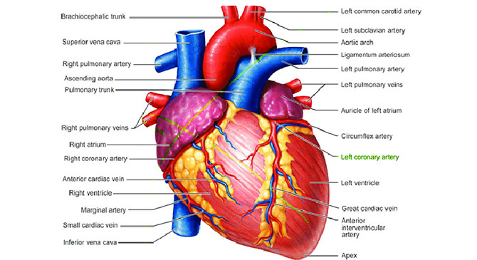
As you can see in the human heart diagram, there are 4 chambers in this tireless pumping organ. All the chambers work in a perfectly coordinated manner for the successful execution of different heart functions. The right chambers contain unclean or deoxygenated blood.
On the other hand, the left chambers contain clean or oxygenated blood. Just look at the different parts of the heart to get a better understanding of the heart's function.
Function of the Blood and Blood Vessels:
You might already know a bit about the function of the blood. Primarily, it serves as a circulatory fluid. The blood carries respiratory gases and nutrients to every individual cell. On its return, it brings carbon dioxide – a waste respiratory gas – for its discharge out of the body.
On the other hand, the function of blood vessels is to serve as a passage for the blood to flow through. The blood vessels are of three types. It is the capillaries where the exchange of gases and nutrients takes place with the individual cells.
Diseases of the Heart
Coronary Artery Disease
The blood vessels that transport blood to the heart become narrow due to plaque deposition. It forces the heart to work harder. As a result, the heart muscle gradually becomes weak. This fatal disease is caused by a high blood cholesterol level.
Myocardial Infarction
It is one of the most dangerous heart diseases. More commonly known as heart attack, myocardial infarction may lead to death on the spot if the individual is unable to get prompt medical help. A heart attack more commonly occurs in patients who are already suffering from coronary artery disease.
The blood flow to the heart is either reduced, or there is a complete blockage. It deprives the heart cells of oxygen. Consequently, all the heart functions come to an end.
Congestive Heart Failure
It is a common heart disease that develops as a result of coronary artery disease or a heart attack. The heart of the patient suffers from damage and is unable to perform the heart function up to its full capacity. As a result, insufficient blood is pumped. And the body's oxygen requirements are not fulfilled. The patients experience fatigue and shortness of breath.
Other Topics Of Heart
- 1. Are the heart and the spleen equal in size?
- 2. Why do the athletes have much larger hearts?
- 3. What prevents impure blood from contaminating impure blood?
- 4. How does the systemic circulation differ from the pulmonary circuit?
- 5.Which heart condition is the most common cause of death worldwide?
- 6. How long does it take to die after the heart stops beating?
- 7. Who believed the heart weighed no more than a feather?
- 8. Is it true about the heart – the bigger the better?
- 9. How can you use your brain to slow down your heartbeat?
- 10. How long does it take the heart to circulate the blood throughout the body?
- 11. How do the hearts of a fetus and an adult differ?
- 12. What is a heart block?
- 13. Why is babies’ heart rate twice as fast as adults?
About the Author